Characterizing Cross Sections and Thin
Structures Using NanoIR Spectroscopy
Home » Characterizing Cross Sections and Thin Structures Using NanoIR Spectroscopy
NanoIR is highly suited for identification of organic components that cannot be conventionally analyzed by other organic analysis techniques such as FTIR or Raman.
Atomic force microscopy-based infrared spectroscopy (AFM-IR, NanoIR) provides vibrational spectroscopic information with high spatial resolution (<30 nm). As a result, NanoIR is highly suited for the identification of organic components in size regimes that cannot be conventionally analyzed by other organic analysis techniques such as FTIR, which is typically limited to tens of microns in analytical area, and even Raman, which is limited to approximately one micron in analytical area.
In this example, two polymer structures were examined: 1) a cross section comprised of three polymers and 2) a thin polymer contaminant on a silicon wafer.
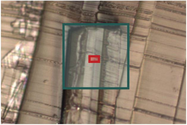
Optical image of the polymer cross section, blue box is the analysis size for FTIR and the red box shows the imaged region in NanoIR.
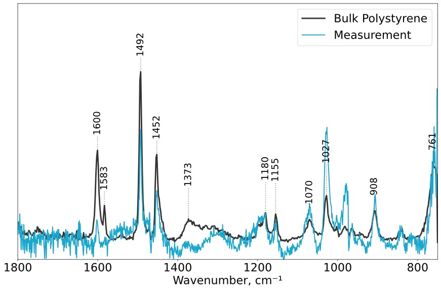
NanoIR spectra taken on the contaminant (blue) and from a bulk film of polystyrene (black).
Contact us today for your NanoIR needs. Please complete the form below to have an EAG expert contact you.
To enable certain features and improve your experience with us, this site stores cookies on your computer.
Please click Continue to provide your authorization and permanently remove this message.