How to interpret TOF-SIMS spectra
Home » How to interpret TOF-SIMS spectra
TOF-SIMS is a technique that can observe elemental, inorganic and molecular species present on the outermost surface of a sample, using a very small primary ion beam dose (~1e12 ions/cm2 or less). It is also quite commonly known as static SIMS because it does not typically induce damage to the sample under investigation. This is in contrast to dynamic SIMS, which causes damage to the sample by creating a crater in the sample over a period of seconds, minutes or hours. . The organic species observed in a TOF-SIMS spectrum may be intact molecular ions or they may be fragment ions created during the sputtering process by the primary ion beam.
The figure below shows the TOF-SIMS spectrum of a common polymer, PET (polyethylene terephthalate, (C10H8O4) n). TOF-SIMS spectra are typically displayed as a plot of signal intensity or ion counts on the Y-axis versus m/z on the X-axis.
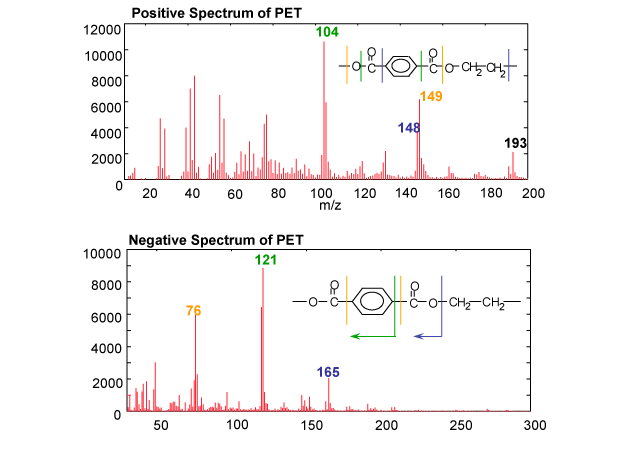
The height of a signal is proportional to the amount of that ion present in the spectrum and the m/z of the ion is basically the mass of that ion (if it is singly charged). From close measurement of the exact mass of the ion, or the difference in m/z between ions, then their molecular formula can be determined. Because n may be a very high number, it is not possible to get intact molecular ions of individual polymer chains into the gas phase using TOF-SIMS. Instead, we observe a series of fragment ions that can provide information about the polymer structure. In this case, a peaked representative of the polymer oligomeric unit can be seen at m/z 193: (C10HO4)H+. Various other peaks are observed in the lower mass range below m/z 193 that are structurally significant and are related to the chemical structure of the sample under analysis. . Careful analysis of the mass and identity of these peaks can enable the polymer structure to be determined, or at least estimated, for complete unknowns. For example, in the case of PET, masses 104, C7H4O+ and 149, C8H5O3+ are detected as prominent fragment ions in the positive ion spectrum, and masses 121, C7H5O2– and 165, C8H4O4– are detected in the negative ion spectrum.. For unknown samples, it is necessary to identify as many of the peaks in the spectra obtained as possible, and from these identified species, piece together the potential identity of the material under investigation.
TOF-SIMS data can also take several other forms:
- The intensity of peaks in spectra can be integrated to allow a relative comparison between samples
- The XY positions of species of interest within the analytical area can be determined and images of the sample created. This allows chemical mapping/imaging of the species present.
- The sample can also be sputtered by operating the primary ion beam or another additional ion beam in a DC mode to erode material (much like dynamic SIMS). After each sputter step spectra or images can be acquired, allowing a depth profile or 3D image to be created.
Would you like to learn more about TOF-SIMS spectra?
Contact us today for your TOF-SIMS spectra needs. Please complete the form below to have an EAG expert contact you.