
In the Lab: Got Cracks?
Join us “In the Lab” to learn how investigative analytical chemistry can help solve your product or manufacturing line problems!

Join us “In the Lab” to learn how investigative analytical chemistry can help solve your product or manufacturing line problems!
In this webinar we introduce the Testing of High Speed I/O to validate the devices from characterization to production
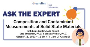
During this live Ask the Expert event, we will answer pre-submitted questions from our audience regarding materials analysis with various X-ray and ion beam analytical techniques.
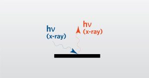
In this webinar we introduce Total Reflection X-ray Fluorescence (TXRF) which is a non-destructive elemental survey technique.
To enable certain features and improve your experience with us, this site stores cookies on your computer. Please click Continue to provide your authorization and permanently remove this message.
To find out more, please see our privacy policy.