Will the Newest Wearable Device Leave You Itching for More Webinar
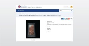
In this webinar we introduce evaluating wearable safety including product recall, lawsuits, and regulatory agency inquiries.

In this webinar we introduce evaluating wearable safety including product recall, lawsuits, and regulatory agency inquiries.

There are many key components of the VCSEL, but one challenging region is the oxide aperture. The oxide aperture is responsible for current confinement, and it is important to have high quality oxidation to prevent failure of the device.
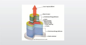
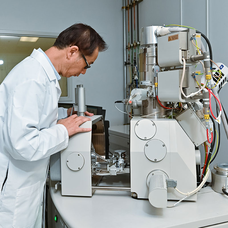
In the full webinar we will introduce analyzing VCSELs with a focus on secondary ion mass spectrometry (SIMS)
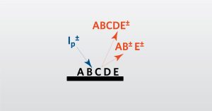
In this webinar we introduce TOF-SIMS which is a surface analysis technique used to investigate the extreme surfaces of samples.
To enable certain features and improve your experience with us, this site stores cookies on your computer. Please click Continue to provide your authorization and permanently remove this message.
To find out more, please see our privacy policy.