sMIM Webinar
In this webinar will be presenting information on scanning Microwave Impedance Microscopy (sMIM) operation.

In this webinar will be presenting information on scanning Microwave Impedance Microscopy (sMIM) operation.

November 2, 2023
Please join us for coffee and conversations! Enjoy a cup of coffee and pastries as you get to know our technical experts.
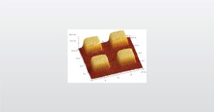
Electrical AFM can measure the electrical/electromechanical properties of various functional materials and samples. This helps distinguish between conductive and nonconductive areas or polar and nonpolar regions of a device.

III-V materials partially provide us with the ever evolving and changing technological advances we enjoy today.
To enable certain features and improve your experience with us, this site stores cookies on your computer. Please click Continue to provide your authorization and permanently remove this message.
To find out more, please see our privacy policy.